PET Scan
Your Affordable PET Scan in Tunisia
Access high-precision diagnosis with a PET Scan: a key exam for detecting or monitoring the progression of cancer, overseen by nuclear medicine specialists.
How does it work?
What is a PET scan?
A PET scan, also known as Positron Emission Tomography, is a nuclear imaging technique used to detect and assess tumors early, determining their size and location. It uses radiopharmaceuticals that emit positron particles. Once injected intravenously, the radiopharmaceutical spreads throughout the patient’s body, thus producing diagnostic images.
What are the indications for a PET scan?
The PET scan in tumor diagnosis
Use a PET scan for better tumor diagnosis and treatment. This method allows you to precisely determine the advancement of cancer and its stage, while avoiding invasive diagnostic procedures. The PET scan is particularly useful for assessing the stage of the tumor, detecting metastases, and monitoring the effectiveness of treatments. In addition, this nuclear imaging technique can also identify and differentiate necrotic areas resulting from radiotherapy.
Who is a PET scan indicated for?
A PET scan is a recommended exam for anyone needing nuclear imaging. It is particularly useful in oncology to assess the stage and effectiveness of therapies, as well as the status of metastases. However, pregnant or breastfeeding women are contraindicated for this exam, as are people who have received radiotherapy less than three months ago or who have undergone surgery or an invasive procedure in the previous month. It is important to mention this information before the exam to facilitate its interpretation. For patients with confirmed diabetes and those undergoing hypoglycemic treatment, a medical evaluation is recommended before the exam.
What is the price of a PET scan in Tunisia?
By choosing to have your PET scan in Tunisia, you benefit from much more affordable prices than those practiced in Europe, while taking advantage of the expertise of our doctors and the modernity of our medical infrastructure. Tunisia Destination Santé offers a PET scan package including all expenses for a 2-night stay in Tunisia. Our packages include the price of your medical stay and all expenses necessary for your PET scan in Tunisia.
Your health, our priority.
Request your free quote.
What is the purpose of a PET scan?
PET scan in the study of tumors
The PET scan plays a crucial role in the diagnosis and management of cancer. Thanks to this technique, it is possible to detect a tumor at an early stage and to determine its extent and possible spread in the body. In addition to this, the PET scan is a valuable tool for accurate cancer staging, allowing the best treatment to be chosen. Finally, it is also essential for evaluating the effectiveness of oncological therapy and for monitoring any recurrence of the tumor after treatment. In short, the PET scan is an essential element in the fight against cancer.
PET scan in neurology
An integral part of the diagnostic process for Alzheimer’s disease and associated disorders, the PET scan can identify areas of the brain with reduced metabolism. In addition, this tool allows for the precise distinction of Alzheimer’s disease from other neurodegenerative disorders.
The PET scan is recommended in neurology to diagnose:
- Epilepsy;
- Cranial trauma;
- Huntington’s disease;
- Stroke;
PET scan in cardiology
PET allows for the assessment of myocardial viability. It helps estimate the benefit that patients suffering from coronary artery disease and ventricular dysfunction can obtain by undergoing a bypass or angioplasty.
PET scan in immunology
- Tuberculous lesions;
- Large vessel vasculitis;
- Sarcoidosis;
- AIDS- or Castleman’s disease-associated infections:
- Potentially infected hepatic or renal cysts;
- Suspected infection with medical devices, such as pacemakers and catheters.
How is a PET scan performed?
Before the PET scan
The PET scan involves administering the radiopharmaceutical intravenously. After waiting for it to enter circulation, the radionuclide leaves a trace: the waiting time to obtain images with the PET varies from about ten minutes to an hour, depending on the radioactive product used.
During the PET scan
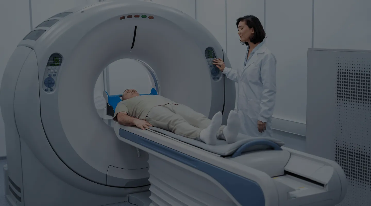
To perform a PET scan, the patient must first lie on a bed and be passed through a ring-shaped machine, about half a meter deep, known as a tomograph. This machine is capable of detecting the radiation emitted by the radiopharmaceutical, while the tomograph records this radiation resulting from the interaction of positrons with the surrounding matter. By rotating at different angles, this device performs precise measurements. The instrument for performing the PET scan thus makes it possible to spatially and temporally reconstruct the biodistribution of the radiopharmaceutical in a given organ or apparatus and to translate it into tomographic images that the doctor interprets.

Do you need a specialist's opinion?
How long does a PET scan take?
A PET scan takes between 20 and 30 minutes, depending on the body part being studied. In total, allow about two to three hours for the entire procedure, from administration of the radiopharmaceutical to the end of the examination.
What are the recommendations for a PET scan?
To prepare for a PET scan, it is necessary to fast for at least 8 hours before the examination. Furthermore, the day before the PET scan, it is strongly recommended to consume abundant amounts of liquids. For good PET preparation, it is generally not recommended to interrupt ongoing medication. It is best to abstain from any strenuous physical activity in the hours before the exam. For diabetic patients and those undergoing hypoglycemic treatment, a medical evaluation before the PET scan is recommended. The PET scan is not painful at all. For optimal use of the radiopharmaceutical product, intravenous administration is required, followed by a 45-minute wait before proceeding with the test. Take advantage of this time to relax and loosen your muscles to ensure optimal results. Once the PET scan is complete, you are free to resume your daily activities normally, although you should take some precautions in the four to five hours following the examination. It is therefore recommended to limit contact with pregnant women and young children until all traces of radioactivity have disappeared from your body. Drinking plenty of water after the PET scan helps to facilitate the elimination of the radioactive product.
Recovery Time After PET Scan
Recovery time after a PET scan is generally short and minimally restrictive. Most patients can resume their normal activities almost immediately after the exam. However, it is recommended to drink plenty of water to help eliminate the radioactive contrast agent used during the procedure. Some patients may experience mild fatigue or minor side effects, such as slight redness at the injection site, but these symptoms quickly disappear. It is also advisable to avoid close contact with pregnant women and young children for a few hours after the scan due to the low radioactivity. In case of any persistent adverse effects, it is important to consult the doctor for appropriate follow-up.
PET Scan Success Rate
The PET scan, or positron emission tomography, is an extremely precise medical imaging tool with a 100% success rate in terms of providing clear and detailed diagnostic images. This technique allows the detection of metabolic abnormalities in tissues and organs, making it particularly effective for the diagnosis and monitoring of diseases such as cancer, heart disease, and neurological disorders. Thanks to the high sensitivity of the PET scan, doctors can obtain crucial information to establish an accurate diagnosis and plan appropriate treatment. However, it is important to note that the success of the PET scan also depends on the patient’s adequate preparation and the expert interpretation of the results by qualified health professionals.
Specifics of PET Scan in Prostate Cancer
The PET scan plays a crucial role in the management of prostate cancer by offering precise visualization of the disease and allowing a detailed assessment of tumor extension. This imaging technique is particularly useful for detecting metastatic lesions that may not be visible on other exams, such as MRI or CT scans. The use of specific markers, such as FDG (fluorodeoxyglucose) or other imaging agents targeting prostate-specific antigen (PSA), allows for the visualization of cancer cells based on their increased metabolism. The PET scan also helps to monitor the response to treatment and detect early recurrences, which is essential for adjusting the therapeutic strategy. In summary, the PET scan is a valuable tool in the diagnosis and management of prostate cancer, offering essential information for informed and personalized decision-making.
Our advantages


Frequently Asked Questions
The PET scan uses radioactive tracers to detect metabolic changes in the brain. This can identify abnormalities associated with diseases like Alzheimer’s, long before symptoms become apparent.
PET scans use positron-emitting tracers, while SPECT scans use gamma-photon-emitting tracers. PET provides higher-resolution images and better accuracy in detecting abnormalities.
By measuring the metabolic activity of cancer cells before and after treatment, a PET scan can show whether the treatment is reducing the activity and size of tumors.
Risks are minimal, as the amount of radiation is low. However, allergic reactions to tracers can occur, although this is rare.
Diabetic patients should monitor their blood sugar before the scan, as high blood sugar levels can affect the results. They should follow their doctor’s specific instructions regarding meals and medications.
Yes, a PET scan can detect infections by identifying areas of inflammation where there is increased metabolic activity.
The PET scan helps study brain function and metabolic abnormalities associated with disorders like schizophrenia and depression, providing crucial information for developing new treatments.
The PET scan is non-invasive and can examine the entire body, detecting possible metastases, while a biopsy is invasive and only examines a tissue sample.
Technological advances have improved image resolution, reduced the amount of radiation needed, and integrated PET with CT scanners for a more comprehensive assessment.
Limitations include limited resolution for small lesions, false positive results due to non-cancerous inflammation, and high costs compared to other imaging techniques.